Learning Objective:
To identify striped muscle cells and nerve cells in animals from prepared slides and draw their labelled diagrams.
Materials Required:

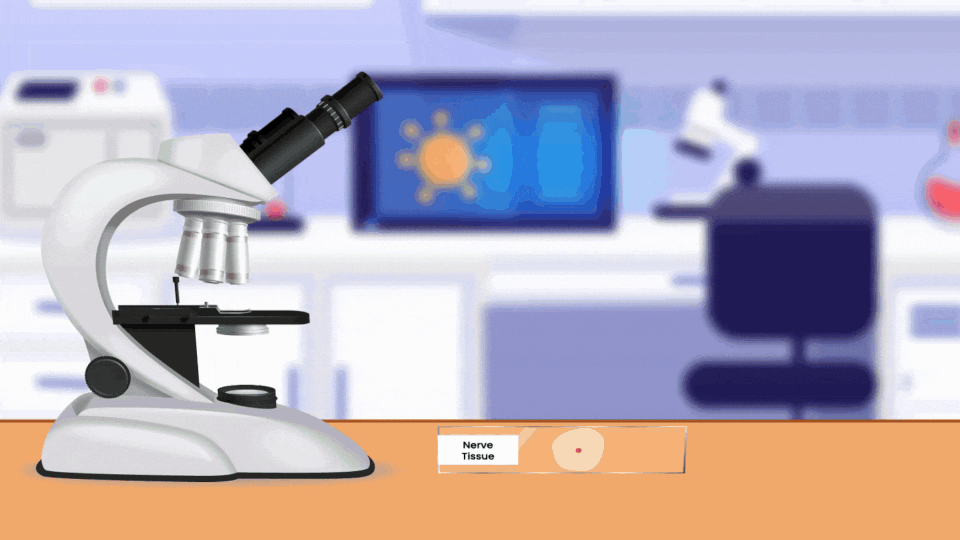
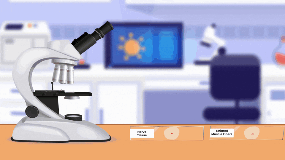
Prepared slides of nerve cells and striated muscle cells
Light/compound microscope
Safety Precautions:
1. HANDLE MICROSCOPE WITH CARE
Experimental Accuracy:
1. Ensure that the microscope is clean
2. Ensure that the most promising region of the slide is in the centre of the stage
3. Ensure that low power is used first, before higher power
Methodology (Optional):
1. Place the prepared slides on the microscope’s stage
2. Observe each slide on low power first, and then high power
3. Sketch diagrams of the tissue in your notebook
Observation:
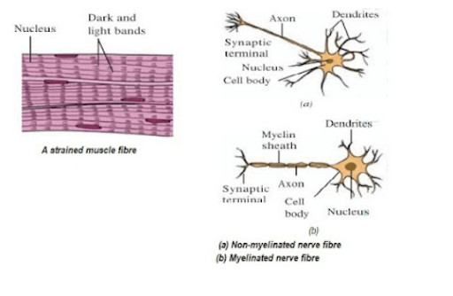
Striated Muscle Fibre:
High Magnification reveals the presence of alternating light and dark bands(which represent thin and thick filaments respectively). The structure observed is multinucleated, as a result of the fusion of muscle cells/fibres(syncytium). The constituent cells are non-tapering, cylindrical and unbranched in nature.
Nerve Cell:
Three distinct sections, those of the dendrites, cell body and axon are seen. The axon may be covered in myelin sheath.
Experiment in Context:
Nerve cells are an integral part of the nervous system. They are constituted by the following fundamental parts:
Axon: conducts electrical impulse(which may trigger the release of neurotransmitter to the synapse)
Schwann's cells: Create myelin sheath at intervals, prevents charge being leaked, and also creating a node of Ranvier, which allows conduction through the gaps instead of the whole axon, making conduction faster, moreover if one part/sheath gets damaged, entire cell won't get damaged
Dendrites: cytoplasmic extensions that connect with other neurons , one of the dendrites extends to form the axon, for conventional neuron diagram, dendrites on soma and on axon's end, allows multiple connections
Nerve cells transfer impulses between two neurons by releasing neurotransmitters across the synapse:
The presynaptic neuron(first neuron) has an electric pulse through the axon that triggers the release of neurotransmitters from vesicles(at the nerve endings/axonic terminals). These neurotransmitters then fuse with the presynaptic membrane. These transmitters then diffuse across the synaptic cleft to the second neuron, binding with its receptors. This triggers an electric impulse to be conducted by the axon of the second neuron. To avoid confusion, synapses only allow one way transmission and destroy neurotransmitters once used so that a pulse isn't transmitted again and again.